ADVANCED STRUCTURAL PROCEDURES
Structural heart disease encompasses conditions that affect the heart’s valves, chambers, or walls, disrupting its ability to pump blood effectively. These conditions can be present at birth (congenital) or develop later in life due to age, other diseases, or infections.
Dr. Fatemi developed the structural heart program at Community Memorial Hospital and St John’s Regional Medical Center. Dr. Fatemi is the Director for Structural Heart at both hospitals. He routinely performs these minimally invasive procedures such as:
These procedures include:
| TAVR WATCHMAN AORTIC VALVULOPLASTY |
MITRAL CLIP (TMVR) PFO CLOSURE ASD CLOSURE |
TAVR – Transcatheter Aortic Valve Replacement
TAVR, or Transcatheter Aortic Valve Replacement, is a minimally invasive procedure to replace a diseased or damaged aortic valve. It’s an alternative to traditional open-heart surgery, offering a less invasive way to treat severe aortic stenosis, a narrowing of the aortic valve. TAVR involves inserting a new artificial valve through a catheter (a thin, flexible tube) into the heart, rather than requiring a large incision in the chest. It’s primarily used for patients with severe aortic stenosis, a condition where the valve narrows, restricting blood flow from the heart. A catheter is inserted into a blood vessel (usually in the groin or chest), guided to the heart, and a new valve is deployed, replacing the diseased valve. TAVR typically involves a shorter hospital stay, less pain, and faster recovery times, while still providing highly effective treatment for severe aortic stenosis. TAVR can be considered for individuals with severe aortic stenosis who are at high risk for open-heart surgery, as well as for individuals who may be at a lower risk.
WATCHMAN
The WATCHMAN procedure is a minimally invasive, one-time procedure to reduce stroke risk in people with atrial fibrillation (AFib), specifically in those with non-valvular AFib. It involves placing a device in the left atrial appendage to close it off, preventing blood clots from forming and potentially traveling to the brain. This procedure is an alternative to lifelong blood thinners and can improve quality of life.
The WATCHMAN device is a small, permanent implant that closes off the left atrial appendage, where blood clots often form in AFib patients. It’s a one-time procedure, meaning it’s not something that needs to be replaced. It’s a minimally invasive procedure, meaning it doesn’t require open-heart surgery.
A catheter is inserted into a blood vessel in the leg and guided to the heart. The device is delivered through the catheter and placed in the left atrial appendage. The device seals off the left atrial appendage, preventing blood clots from forming and traveling to the brain.
WHO QUALIFIES: People with non-valvular AFib, meaning their AFib is not caused by a heart valve problem. Those who are at risk of stroke due to AFib. Patients who are currently taking or have considered taking blood thinners like warfarin. Individuals who have a high risk of bleeding while on blood thinners.
The Watchman procedure reduces the risk of stroke in AFib patients. It can eliminate the need for lifelong blood thinners, such as warfarin, improve quality of life by reducing the need for blood tests and dietary restrictions associated with blood thinners.
During the Watchman procedure general anesthesia is administered. A small incision is made in the leg (usually the groin) to access a blood vessel. A catheter is inserted into the blood vessel and guided to the heart. The WATCHMAN device is delivered through the catheter and placed in the left atrial appendage. The catheter is removed, and the incision is closed.
The procedure typically takes about an hour. Patients usually stay in the hospital overnight.
AORTIC VALVULOPLASTY
Aortic Valvuloplasty also known as balloon aortic valvotomy is the widening of a stenotic aortic valve using a balloon catheter inside the valve. The balloon is placed into the aortic valve that has become stiff from calcium buildup.
MITRAL CLIP (TMVR)
A Mitral Valve Clip, also known as TMVR (Transcatheter Mitral Valve Repair) or MitraClip, is a minimally invasive procedure used to repair a leaky mitral valve. It addresses mitral regurgitation, where blood flows backward through the valve instead of forward. The procedure uses a small metal clip, delivered via a catheter inserted into a blood vessel in the leg, to grasp and partially close the abnormal valve leaflets, reducing or eliminating the leak.
WHAT IS MITRAL VALVE REGURGITATION?
The mitral valve is located between the heart’s two left chambers and has two leaflets that open and close to allow blood flow.
Mitral regurgitation occurs when the mitral valve doesn’t close properly, allowing blood to leak backward.
This can cause symptoms like shortness of breath, fatigue, and swelling in the legs.
HOW DOES THE MITRAL VALVE CLIP WORK?
Minimally Invasive: The procedure is performed without open-heart surgery, meaning there’s no incision in the chest.
Catheter Delivery: A small metal clip is inserted through a catheter, which is guided to the mitral valve through a blood vessel in the leg.
Leaflet Attachment: The clip is attached to the edges of the defective valve leaflets, holding them together to partially close the valve.
Reduced Backflow: This helps to restore normal blood flow and improve heart function.
WHO IS A CANDIDATE FOR A MITRAL VALVE CLIP?
Patients with mitral regurgitation who are at high risk for or unable to undergo open-heart surgery.
Patients whose symptoms are severe or not adequately controlled by medication.
BENEFITS OF A MITRAL VALVE CLIP:
Minimally invasive, reducing the risk of complications and recovery time.
It can significantly improve symptoms like shortness of breath and fatigue.
In summary, the mitral valve clip is a valuable treatment option for patients with mitral regurgitation who are at high risk for open-heart surgery or who have not responded well to other treatments.
PFO CLOSURE
Percutaneous closure is a surgical procedure used to treat patients with patent foramen ovale (PFO) and atrial septal defect (ASD). Advancements in device technology and image guidance now permit the safe and effective catheter-based closure of numerous intracardiac defects, including PFO and ASD.
Dr. Omid Fatemi performs PFO Closures at Community Memorial Hospital.
ASD CLOSURE
ASD CLOSURE – Atrial Septal Defect (ASD) is an opening or hole in the wall that separates the two upper chambers of the heart. This wall is called the atrial septum. The hole causes oxygen-rich blood to leak from the left side of the heart to the right side. Different types of closure devices are used to close a hole or an opening between the right and left sides of the heart. Some of these birth defects are located in the wall (septum) between the upper chambers (atria) of the heart: Patent Foramen Ovale (PFO) Atrial Septal Defect (ASD).
Dr. Omid Fatemi performs these ASD Closures at Community Memorial Hospital.
ADVANCED ELECTROPHYSIOLOGY PROCEDURES
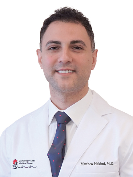
Dr. Jonathan Dukes, and Dr. Matthew Hakimi, Cardiac Electrophysiology Physicians, provide the most advanced evaluations, treatments and procedures for atrial fibrillation (AFib) and other circuitry issues to Ventura County residents. Dr. Dukes is the Director of Electrophysiology at Community Memorial Hospital.
Dr. Jonathan Dukes, and Dr. Matthew Hakimi specialize in complex ablations and are accepting new patients for first-time evaluations and new patients seeking to have a redo ablation.
Advanced Electrophysiology procedures include:
| WATCHMAN ABLATION AFib ABLATION ATRIAL FLUTTER ABLATION |
SVT ABLATION VT ABLATION PVC (Premature Ventricular Contraction) PULSE FIELD ABLATION |
WATCHMAN
The WATCHMAN procedure is a minimally invasive, one-time procedure to reduce stroke risk in people with atrial fibrillation (AFib), specifically in those with non-valvular AFib. It involves placing a device in the left atrial appendage to close it off, preventing blood clots from forming and potentially traveling to the brain. This procedure is an alternative to lifelong blood thinners and can improve quality of life.
The WATCHMAN device is a small, permanent implant that closes off the left atrial appendage, where blood clots often form in AFib patients. It’s a one-time procedure, meaning it’s not something that needs to be replaced. It’s a minimally invasive procedure, meaning it doesn’t require open-heart surgery.
A catheter is inserted into a blood vessel in the leg and guided to the heart. The device is delivered through the catheter and placed in the left atrial appendage. The device seals off the left atrial appendage, preventing blood clots from forming and traveling to the brain.
Who qualifies: People with non-valvular AFib, meaning their AFib is not caused by a heart valve problem. Those who are at risk of stroke due to AFib. Patients who are currently taking or have considered taking blood thinners like warfarin. Individuals who have a high risk of bleeding while on blood thinners.
The Watchman procedure reduces the risk of stroke in AFib patients. It can eliminate the need for lifelong blood thinners, such as warfarin, improve quality of life by reducing the need for blood tests and dietary restrictions associated with blood thinners.
During the Watchman procedure general anesthesia is administered. A small incision is made in the leg (usually the groin) to access a blood vessel. A catheter is inserted into the blood vessel and guided to the heart. The WATCHMAN device is delivered through the catheter and placed in the left atrial appendage. The catheter is removed, and the incision is closed.
The procedure typically takes about an hour. Patients usually stay in the hospital overnight.
ABLATION
AFib ABLATION
ATRIAL FLUTTER ABLATION
SVT ABLATION
VT ABLATION
PVC (Premature Ventricular Contraction)
PULSE FIELD ABLATION


