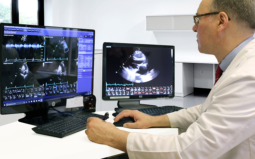
ADULT ECHOCARDIOGRAPHY
Adult Echocardiography (Echocardiogram) is a diagnostic test that uses ultrasound waves to make images of the heart chambers, valves, and surrounding structures. It can measure cardiac output and is a sensitive test for fluid around the heart (pericardial effusion).
Dr. Brackett and Dr. Mansour hold the distinction of adult echocardiography testamur. This requires the completion of a special competence examination to optimize skill in the performance and interpretation of cardiac ultrasound. Cardiology Associates echo lab is accredited with ICAEL.
BUBBLE STUDY ECHOCARDIOGRAM
CAROTID ULTRASOUND
ARTERIAL ULTRASOUND
ABDOMINAL ULTRASOUND
RENAL ULTRASOUND (KIDNEY)
VENOUS DOPPLER ULTRASOUND
STRESS ECHOCARDIOGRAM TEST

NUCLEAR STRESS TEST – MYOCARDIAL PERFUSION STUDY
A Myocardial Perfusion Study, also known as a Nuclear Stress Test, is a non-invasive imaging test that assesses blood flow to the heart muscle (myocardium). This test may be done with or without exercise. It helps diagnose and assess conditions like coronary artery disease, heart damage from heart attacks, and blood flow patterns.
A small amount of a radioactive tracer is injected into a vein, and a special camera (gamma camera) is used to detect the tracer’s distribution in the heart. The tracer will be absorbed by healthy heart tissue, and areas with poor blood flow will show up as darker areas on the scan.
Dr. Steinberg and Dr. Patel are certified in nuclear cardiology tests.
![]()
NUCLEAR PET SCAN (Positron Emission Tomography)

A nuclear PET (Positron Emission Tomography) scan of the heart is a non-invasive imaging test that uses radioactive tracers to visualize the heart and its blood flow, helping doctors diagnose and assess heart conditions. It provides detailed 3D images of the heart’s activity, showing how well the heart muscle is working and where blood flow may be restricted. A nuclear PET scan is the fastest, most accurate way to detect heart disease and any abnormalities that could possibly have the potential for a heart attack. This test may also help your cardiologist to devise a more specific treatment plan for patients with known coronary artery disease or heart attack.
If you are scheduled for a nuclear PET scan, you will be given detailed instructions in preparation for your test, and during the testing process. If you have any further questions, please contact our offices at 805-278-4020.
CARDIOVASCULAR COMPUTED TOMOGRAPHY (CARDIAC CT)

Dr. Alon Steinberg is certified to interpret Cardiac CT scans.
HOSPITAL PROCEDURES
HEART CATHETERIZATION
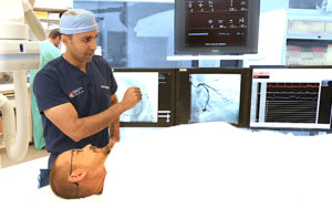 |
Cardiac Catheterization is a procedure used to diagnose and treat cardiovascular conditions. It is performed as an outpatient procedure in the hospital. During cardiac catheterization, a long thin tube called a catheter is inserted in an artery or vein in the groin, neck or arm (transradial) and threaded through your blood vessels to the heart to determine if there is disease of the heart muscle, valves or coronary (heart) arteries. During the procedure, the pressure and blood flow in the heart can be measured. |
PERCUTANEOUS CORONARY INTERVENTION (PCI)
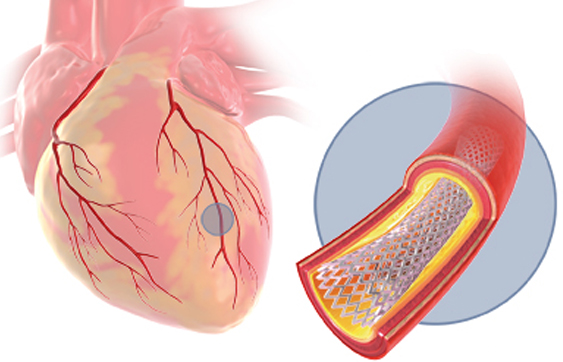 |
During a cardiac catheterization if it is determined that there are blockages that would benefit from a coronary stent, then a small tube-shaped device (stent) will be placed via the already in-place catheter, to keep the blocked coronary arteries open and increasing blood flow to the heart.
Dr. Shaun Patel, Dr. Omid Fatemi and Dr. Sogomonian are Interventional Cardiologists at Cardiology Associates. |
CTO PERCUTANEOUS CORONARY INTERVENTION
Dr. Omid Fatemi specializes in this procedure.

Dr. Fatemi performs more advanced procedures within our structural heart program. Dr. Fatemi is the Director for the Structural Heart Department at Community Memorial Hospital and St. John’s Regional Medical Center.
These procedures include:
| TAVR WATCHMAN AORTIC VALVULOPLASTY |
MITRAL CLIP PFO CLOSURE ASD CLOSURE |
How Structural Heart Expertise Addresses Heart Issues with Dr. Omid Fatemi Omid Fatemi, MD
ELECTROPHYSIOLOGY

Dr. Jonathan Dukes, Cardiac Electrophysiology Physician, provides the most advanced evaluations, treatments and procedures for atrial fibrillation (AFib) and other circuitry issues to Ventura County residents. Dr. Dukes is the Director of Electrophysiology at Community Memorial Hospital.
Dr. Jonathan Dukes performs pacemaker and defibrillator implantations.
Dr. Shaun Patel also performs pacemaker implantations.
ELECTROPHYSIOLOGY TESTING
EVENT MONITORING
TILT TABLE TEST
ELECTROPHYSIOLOGY PROCEDURES
PACEMAKER IMPLANTATION
A pacemaker insertion is the implantation of a small electronic device that is usually placed in the chest (just below the collarbone) to help regulate slow electrical problems of the heart. A pacemaker may be recommended to ensure that the heartbeat does not slow to a dangerously low rate.
DEFIBILLATOR IMPLANTATION
An implantable cardioverter defibrillator (ICD) is a small, battery-powered device placed under the skin, typically in the chest, to monitor and correct life-threatening heart rhythm problems (arrhythmias). It can deliver electrical shocks to restore a normal heart rhythm when needed.
PACEMAKER AND DEFIBRILLATOR BATTERY REPLACEMENT
Battery replacement depends on how often your system uses the device. You will be advised about your battery life during routine device monitoring.
LEADLESS PACEMAKER
A leadless pacemaker is an implantable pulse generator (IPG), an implanted medical device that restores your heart rate to a more normal rhythm by stimulating the heart with precisely timed pulses of electricity. Unlike most pacemakers that have leads that go into heart, leadless pacemakers are implanted directly into the heart and have no leads.
CARDIAC RESYNCHRONIZATION THERAPY (CRT) DEVICES
CRT stands for cardiac resynchronization therapy. A CRT device is a pacemaker or defibrillator that sends small electrical impulses through leads to one upper and the two lower chambers of your heart. You don’t feel them, but they help all three chambers beat in sync. This makes it easier for your heart to pump blood and oxygen throughout your body.
IMPLANTABLE MONITORING DEVICE
Reveal LINQ, Insertable Cardiac Monitor, is the world’s smallest implantable cardiac monitoring device. The procedure to implant one just takes a few minutes to perform as an outpatient. It records the heart rhythm continuously to capsure recurrent unexplained episodes of palpitations.
CATHETER ABLATION
Dr. Jonathan Dukes, Cardiac Electrophysiology Physician, provides the most advanced evaluations, treatments and procedures for atrial fibrillation (AFib) and other circuitry issues to Ventura County residents. Dr. Dukes is the Director of Electrophysiology at Community Memorial Hospital.
Dr. Jonathan Dukes specializes in complex ablations and are accepting new patients for first-time evaluations and new patients seeking to have a redo ablation.
Advanced Electrophysiology procedures include:
| WATCHMAN ABLATION AFib ABLATION ATRIAL FLUTTER ABLATION |
SVT ABLATION VT ABLATION PVC (Premature Ventricular Contraction) PULSE FIELD ABLATION |
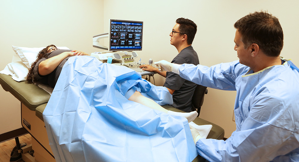
PERIPHERAL VASCULAR PROCEDURES


PERIPHERAL ANGIOGRAM
ENDOVASCULAR PERCUTANEOUS ANGIOPLASTY INTERVENTION
Endovascular Percutaneous Angioplasty Intervention – Endovenous percutaneous angioplasty, also known as percutaneous transluminal angioplasty (PTA) or endovascular angioplasty, is a minimally invasive procedure used to open blocked or narrowed veins. It involves inserting a thin tube (catheter) with a balloon on its tip into the vein, inflating the balloon to widen the vessel, and sometimes placing a stent (a small mesh tube) to keep it open. Endovascular percutaneous angioplasty intervention may include transfemoral carotid artery, subclavian, and lower extremity for claudication.
ENDOVASCULAR ABDOMINAL AORTIC ANEURYSM (AAA) REPAIR
Endovascular Abdominal Aortic Aneurysm (AAA) Repair, or EVAR, is a minimally invasive surgical procedure to repair weakened areas in the aorta, the large blood vessel that carries blood from the heart to the rest of the body. It involves inserting a stent through small incisions in the groin, guided by X-rays, to reinforce the aorta and prevent the aneurysm from bursting.
SUBCLAVIAN VEIN STENTING
Subclavian vein stenting is a minimally invasive procedure used to treat narrowing or blockage (stenosis) in the subclavian vein, a major vein in the upper body that carries blood back to the heart. Stents are small, expandable mesh tubes that are inserted into the vein to open it up and restore proper blood flow. This procedure is often performed to address conditions like venous thoracic outlet syndrome or to manage blockages caused by central lines or other medical devices.
Venous disease, deep and superficial veins including iliac vein stenting and venous thrombectomy for deep vein thrombosis
ILIAC VEIN STENTING
Iliac vein stenting is a procedure that uses a stent, a small mesh tube, to open and maintain the patency of the iliac veins, which are in the pelvis. This procedure is often performed to treat iliac vein compression, also known as May-Thurner syndrome and other conditions that lead to chronic venous insufficiency, such as pelvic congestion syndrome.
VENOUS THROMBECTOMY
Venous thrombectomy is a surgical procedure used to remove a blood clot (thrombus) from a vein, typically used to treat deep vein thrombosis (DVT) when other treatments aren’t effective. It can be performed percutaneously.
PULMONARY EMBOLISM TREATMENT
Pulmonary embolism treatment, including mechanical thrombectomy and catheter-directed thrombolysis
Varicose Veins Treatment
Endovenous Ablation is an image-guided, minimally invasive treatment for varicose veins. It uses radiofrequency or laser energy to cauterize (burn) and close the varicose veins. The results are remarkable. Patients who suffer with painful, disfiguring varicose veins now have the opportunity for a simple procedure with minimal pain.
Our vein lab founded in 2009, is located in Oxnard, CA. We treat patients who meet medically based requirements. Vein ablations are covered by most insurances.
Dr. Shaun Patel and Dr. Robert Sogomonian are board-certified in endovascular procedures.
